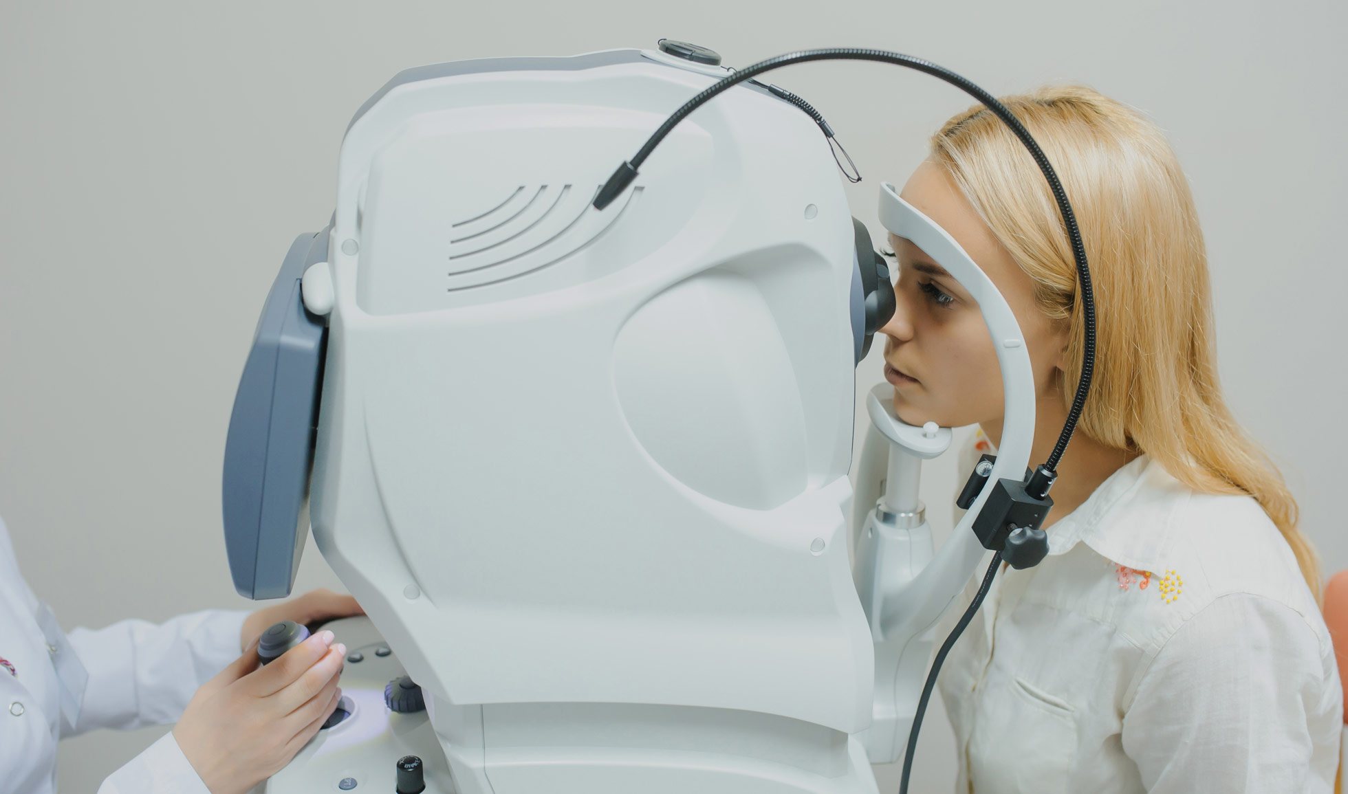
An eye exam involves a series of tests to evaluate your vision and check for eye diseases. Your eye doctor is likely to use various instruments, shine bright lights at your eyes and request that you look through an array of lenses. Each test during an eye exam evaluates a different aspect of your vision or eye health.
This test evaluates the muscles that control eye movement. Your eye doctor watches as your eyes follow a moving object, such as a pen or small light. Afterwards, he looks for muscle weakness, poor control or poor coordination.
This test measures how clearly you see. Your doctor asks you to identify different letters of the alphabet printed on a chart or a screen positioned some distance away.
Each eye is tested separately. Your near vision also may be tested, using a card with letters held at reading distance.
Light waves are bent as they pass through your cornea and lens. If light rays don’t focus perfectly on the back of your eye, you have a refractive error. That can mean you need some form of correction, such as glasses, contact lenses or refractive surgery, to see as clearly as possible.
Assessment of your refractive error helps your doctor determine a lens prescription that will give you the sharpest,
most comfortable vision. The assessment can also determine that you don’t need corrective lenses.
Your doctor may use a computerized refractor to estimate your prescription for glasses or contact lenses. Or he or she may use a technique called retinoscopy. In this procedure, the doctor shines a light into your eye and measures the refractive error by evaluating the movement of the light reflected by your retina back through your pupil.
Your eye doctor usually fine-tunes this refraction assessment by having you look through a masklike device that contains wheels of different lenses (phoropter). Afterwards, he asks you to judge which combination of lenses gives you the sharpest vision.
A slit lamp is a microscope that magnifies and illuminates the front of your eye with an intense line of light. Your doctor uses
this device to examine the eyelids, lashes, cornea, iris, lens and fluid chamber between your cornea and iris.
Your doctor may use a dye, most commonly fluorescein, to color the film of tears over your eye. This helps reveal damaged cells on the front of your eye. Your tears wash the dye from the surface of your eye fairly quickly.
This examination — sometimes called ophthalmoscopy or fundoscopy — allows your doctor to evaluate the back of your eye, including the retina, the optic disk and the retinal blood vessels that nourish the retina. Having your pupils dilated with eyedrops before the exam keeps your pupils from getting smaller when your doctor shines light into the eye.
After administering eyedrops and giving them time to work, your eye doctor may use one or more of these techniques to view the back of your eye:
Direct exam. Your eye doctor uses an ophthalmoscope to shine a beam of light through your pupil to see the back of the eye. Sometimes eyedrops aren’t necessary to dilate your eyes before this exam.
Indirect exam. During this exam, you might sit up or be reclined in the exam chair. Your eye doctor examines the inside of the eye with the aid of a condensing lens and a bright light mounted on his or her forehead. This exam lets your doctor see the retina and other structures inside your eye in great detail and in three dimensions.
Whether you need vision correction, either through glasses, contact lenses or surgery
Whether your eyes are healthy, or you have cataracts, glaucoma or retinal disorders, such as macular degeneration or diabetic retinopathy.
If you need corrective lenses, your doctor will give you a prescription. If your eye exam yields other abnormal results, your doctor will discuss with you the next steps for further testing or for treating an underlying condition.