Corneal Topography
 Athens EyeCare Clinicathenseyecare.gr · (+30) 210 777 8515
On this page
What is corneal topography?
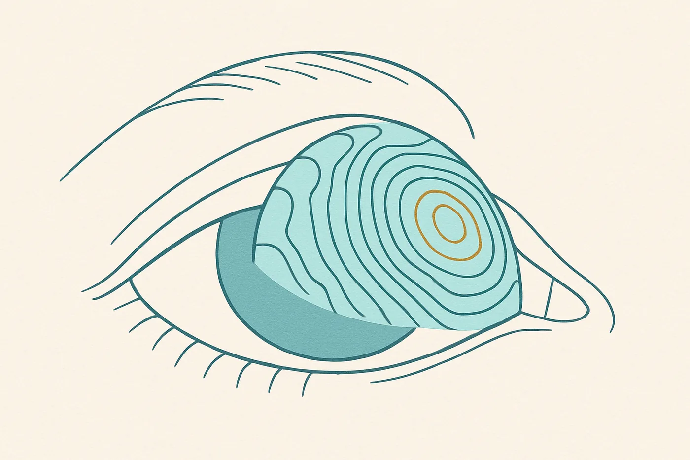
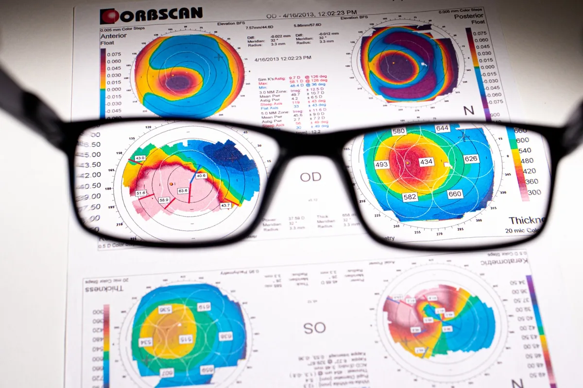
Corneal topography is a specialised photographic technique that maps the surface of the cornea, the clear front window of the eye. A topography scan allows the doctor to detect distortions in the curvature of the cornea, which is normally smooth. It also helps doctors monitor eye disease and plan surgery.
Which conditions is corneal topography used for?
Scarring
Trauma (injury) or infection can scar the cornea, changing its shape. A topography scan measures the distortion and its effect on vision.
Growths
The size of pterygia and other growths can be monitored with topography.
Astigmatism and keratoconus
Topography can help detect astigmatism and early keratoconus, and track how they progress.
Contact lens fitting
Topography scans help determine which type of contact lens will best improve vision. If a scan shows significant distortion, a special rigid contact lens (RGP) can sometimes correct it.
How does corneal topography help with surgery?
Refractive surgery
In refractive surgery such as LASIK, the shape of the cornea is altered to correct refractive errors such as myopia. A topography scan helps the surgeon plan precisely how to reshape the cornea.
Cataracts
When a cataract clouds the eye’s natural lens, the lens is replaced with an intraocular lens (IOL) during cataract surgery. In some cases, corneal topography helps the surgeon select the right IOL.
Corneal transplants
After a corneal transplant, the surgeon may use corneal topography to help the eye heal correctly. Based on the shape of the cornea, the images help determine which stitches should be removed, and when.
Corneal cross-linking
Corneal cross-linking is a procedure that strengthens a cornea affected by keratoconus. A topography scan may be performed to see whether this treatment is needed, and further scans are taken afterwards to monitor the eye.
What to expect during a corneal topography scan
-
You will be seated facing a large bowl with illuminated circles inside it. Chin and forehead rests keep your head steady, so the clearest possible images can be captured.
-
You will be asked to stare at a fixed target inside the bowl while the pictures are taken.
-
The scan takes only a few seconds, though it may need to be repeated a few times.
-
Corneal topography is painless, as nothing touches your eye during the scan.
Athens EyeCare Clinic uses one of the most advanced corneal topographers in the world, the Pentacam HR from Oculus, which provides reliable and accurate measurements.