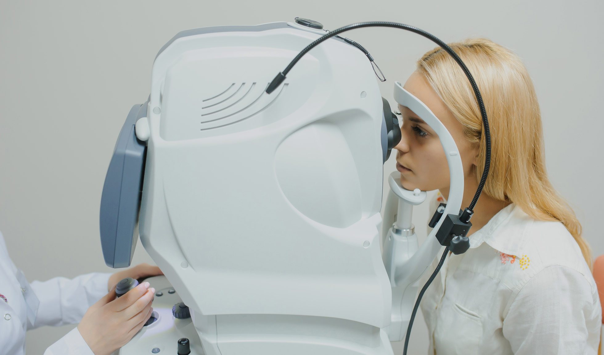
Fundus photography is the process of taking serial photographs of the interior of your eye through the pupil. A fundus camera is a specialized low-power microscope attached to a camera used to examine structures such as the optic disc, retina and lens.
Before the procedure, your ophthalmologist may administer an eye drop to increase the size of the pupil. This allows your doctor to examine the interior surface of your eye much more properly.
Your doctor will instruct you to sit in front of the fundus camera with your chin on a chin rest (an attachment) and your forehead against the bar.
Your doctor will focus and align the fundus camera on the pupil (the black center of your eye).
As soon as the doctor presses the shutter release, flash fires that create a photograph of the interior surface of your eye.
The benefits include:
It is a non-invasive procedure and only takes a minute or two.
It is easier to visualize the details of the retina (the interior surface of the eye) in stereoscopic fundus photographs rather than examining your eye directly.
It provides a bird’s eye view of entire layers on the retina (the interior surface of the eye) and allows your doctor to provide the most accurate diagnosis.
Many times, certain internal eye landmarks are more visible in fundus photography than other eye examinations, for example, fluorescein angiography (an eye examination in which a fluorescent dye is injected inside your eye).
Serial photographs of the internal structure of your eye may allow your doctor to study the delicate changes in the eye nerve, and they can recommend the appropriate therapy to you.
It allows early and accurate diagnosis, especially changes in the eye of patients with diabetes and blood pressure, which are essential for timely treatment and improvement in the patient’s outcome.
You get pictures of the current appearance of the retina (the interior surface of your eye) that is more worthy sometimes than the physician’s notes.
Athens Eye Care Clinic has one of the most modern fundus cameras with Autofluorescence, Digital Fluoroangiography and Digital Indocyanine Angiography, the Visucam 524 from the Zeiss company.