Digital Anterior Segment Photography

What is an Digital Anterior Segment Photography;

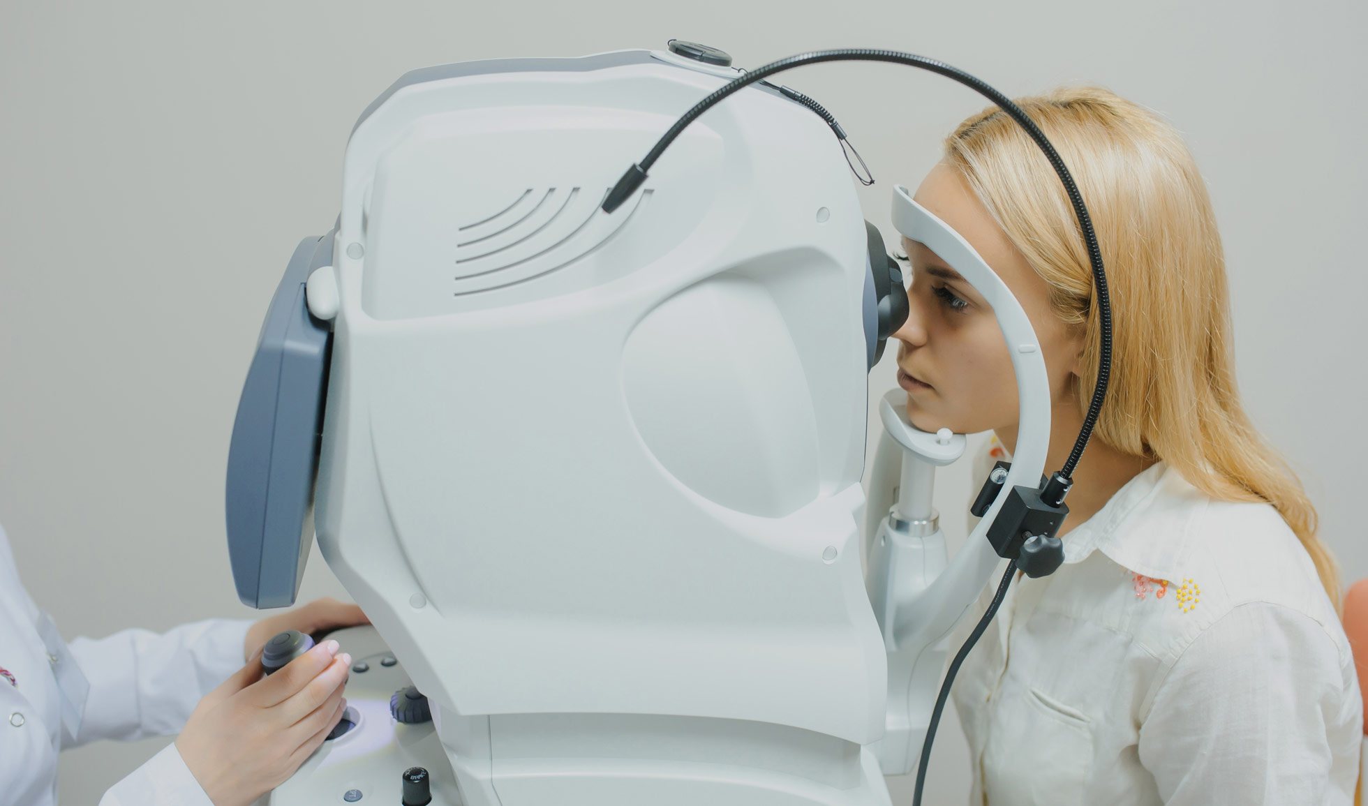
Anterior segment and external imaging involves a series of images being taken of the front of your eye and around the eye.

Anterior segment and external imaging involves a series of images being taken of the front of your eye and around the eye.

The test is carried out to help document the appearance of the cornea, conjunctiva, iris and other structures that are at the front of the eye and around the eye. It is also used to monitor certain conditions.

The images will help the eye doctor in monitoring your condition or may be used to help diagnose a condition.

The imaging may include the use of a bright light or flash. You will find that your vision may be a strange colour after the imaging, but this does not last long.

It may be possible, under certain circumstances, to use
different equipment to image the front of your eye. If you have any concerns, please discuss these with the doctor and they will go through which options are available to you.

You may need to have drops put in your eyes to dilate your pupils. It will take 20-30 minutes for your pupils to fully dilate. The drops can

make your vision blurred for 2 to 24 hours and so it is recommended that you do not drive. It is therefore important that you make appropriate transport arrangements.

You will be asked to sit at an instrument similar to the one the doctor has used to examine your eye. The imaging normally takes between 10 and 20 minutes.

Some patients will need to have an examination with a contact lens. If this is required, local anaesthetic drops will be used to numb the front of the eye. The drops will last for about 20 minutes.

Some patients will need to have a special-coloured eye drop added prior to imaging. This will not change your vision or have any long-lasting effect.


Athens EyeCare clinic has one of the most advanced anterior segment photography, organize an appointment and see it yourself.

All the Diagnostic Examinations